


Dr. Sathyasree Viswanathan
Institution: Apollo Cancer Institute, Chennai
Carcinoma left breast(1975), treated and breast implantation done. Implant rupture (2012). Follow up mammogram in 2017, shows left axillary adenopathy.
Discussion
Rupture of a silicone breast implant can be either intracapsular or extracapsular. The latter, while less common can extravasate into surrounding breast tissue, lung parenchyma, chest-wall muscles and lymph nodes in the axilla and supraclavicular fossa. Subcutaneous siliconomas in more distal areas such as the abdominal wall, inguinal region and lower limbs can also occur. Pathology: Silicon induces a foriegn body reaction with giant cell formation and phagocytosis leading to the formation of a granuloma called ‘siliconoma’. This occurs in extracapsular rupture due to “silicone bleeding” through the membrane of the prosthesis and the fibrous capsule. Other complications including painful nodularity, skin erosions, migration along tissue planes or distant locations with embolism and even death. Radiographic features: Mamography: Extremely dense lobulated masses with or without rim calcification. Extremely dense lymph nodes may be present. Ultrasound: Free silicone can appear as hypoechoic masses which are almost indistinguishable from cysts and are usually surrounded by echogenic noise. CT: Free silicone presents as isodense nodulations and subcutaneous involvement with infiltrative, nodular or confluent pattern and lymphnodal involvement. MRI: MRI shows silicone nodules and adenopathies with high signal intensity on T2- weighted images (although less than the water) and silicone-emphasised sequences and low signal intensity on silicone-suppressed sequences.
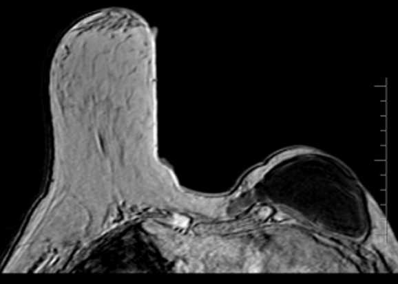
Findings
MAMMOGRAM and USG: Enlarged right axillary nodes MRI: Extra capsular rupture of silicone implant along the medial aspect with minimal leak of silicone gel into the anterior chest wall and adjacent breast parenchyma. PET-CT: Hypermetabolic soft tissue deposits in left anterior chest wall and right rectus abdominis. Hypermetabolic regional and distant nodes.
Answer
Diagnosis: Disseminated siliconomas
References
Imaging findings of mammary and systemic silicone deposition secondary to breast implants Naziya Samreen, Katrina N Glazebrook, Asha Bhatt, Sudhakar K Venkatesh, Brendan P McMenomy, Anupam Chandra, Shuai leng, Kalie E Adler, and Cynthia H McCollough The British Journal of Radiology 2018 91:1089
Together we are making a difference- and, you can too..
Breast Imaging Society, India was founded by a group of dedicated breast radiologists in 2013 with the idea to improvise and standardize breast imaging in India.