Findings
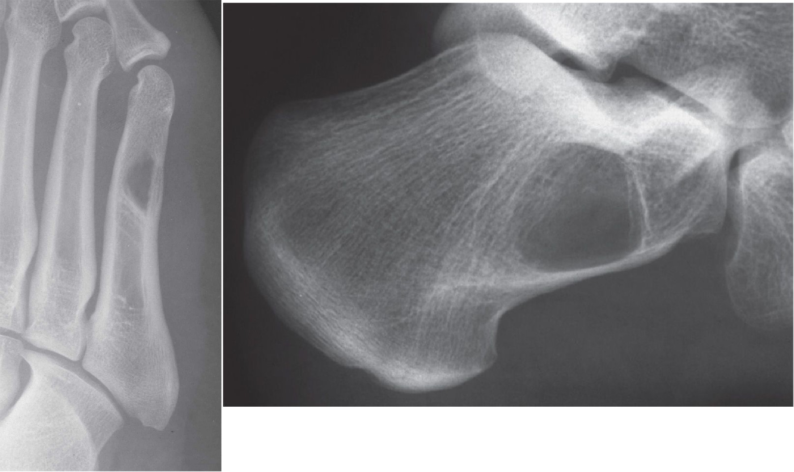
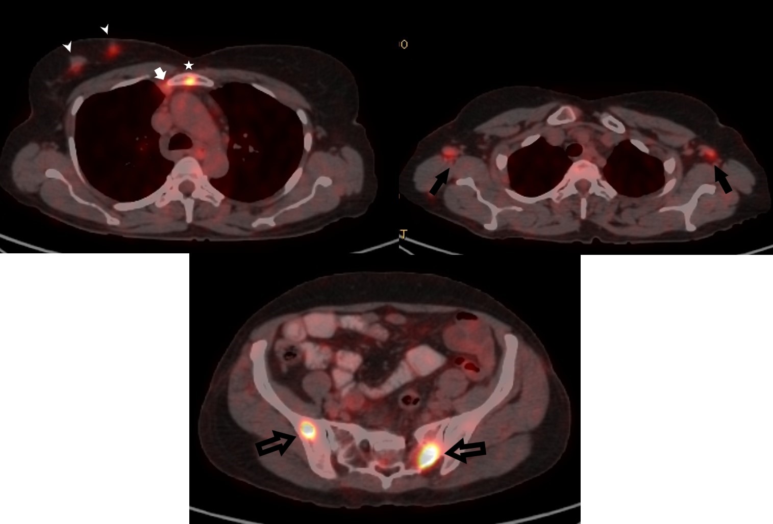
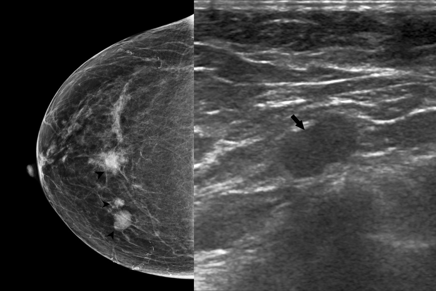
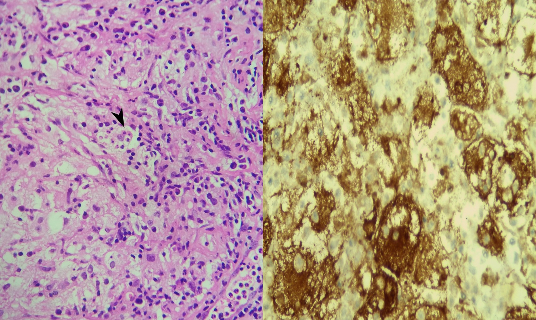
Findings Extremity X rays taken at the time of presentation to revealed lytic lesions in the metatarsal and the calcaneum. Subsequently she underwent a PET CT which revealed multifocal right breast lesions in the upper inner quadrant (White arrow heads) with right internal mammary node (white arrow) and sternal lesion (white star ).Bilateral axillary nodal uptake(solid black arrows)was seen along with multiple other lytic bony lesions (block black arrows ) Pre -biopsy Right mammogram (RCC) showed a larger spiculated and smaller irregular masses in the upper inner quadrant (Black arrow heads) .Axillary nodes showed diffuse cortical thickening on USG (black arrow),which were concerning for metastasis.Core biopsy specimen stained with H&E (x 400) demonstrated diffuse dense infiltration by lymphocytes, plasma cells and histiocytes. Emperipolesis- meaning phagocytosis of lymphocytes by histiocytes (Black arrow head) was seen in the samples.Immunohistochemistry revealed histiocytes demonstrating positivity for S100(x 400)
Answer
Answer :Extra-nodal Rosai Dorfman Disease of the Breast Differential Diagnosis :Multifocal breast malignacy,lymphoma,metastasis
Discussion
Discussion: In 1969 Rosai and Dorfman established sinus histiocytosis with massive lymphadenopathy (SHML) as a benign clinicopathologicentity (1).The disease more popularly known as Rosai- Dorfman disease has a worldwide distribution with approximately 80% cases being detected in the first two decades. Though the exact etiopathogenesis remains unclear, viral agents like EBV and herpes have been implicated.Most commonly patients present with bilateral painless cervical lymphadenopathy which tend to coalesce over time to form multinodular masses as disease progresses. Rosai-Dorfman disease is a rare disorder wherein there is excessive production and accumulation of histiocytes in the lymph nodes. This excessive deposition may occur in other areas like the skin, central nervous system, kidney, and digestive tract. Also referred to as extra nodal RDD these may mimic other diseases in these organs. Extra nodal RDD may be seen in as high as 43% cases and commonly affects the skin, upper respiratory track and bones (2). Rosai-Dorfman disease of the breast is a rare benign inflammatory disorder that can mimic breast cancer clinically and on imaging studies. In the breast RDD usually presents as a painless, palpable mass lesion seen incidentally or on screening mammography.They have been usually described as obscured or even spiculated lesions.Larger lesions tend to infiltrate the surrounding breast parenchyma resulting in a echogenic halo mimicking desmoplasia seen in breast malignancies.These lesions tend to be vascular. Akin to other benign and malignant lymphoproliferative disorders, RDD also shows FDG –avidness. This is due to the glucose dependence of the inflammatory cells and the proliferating histiocytes (3) This case highlights how RDD mimicked a metastatic breast malignancy on nuclear imaging due to the presence of FDG –avid lesions in the breast, loco-regional nodes and in the bones.
Reference
1.Pham CB, Abruzzo LV, Cook E, Whitman GJ, Stephens TW (2005) Rosai-Dorfman disease of the breast. AJR Am J Roentgenol 185: 971-972. 2. Foucar E, Rosai J, Dorfman R (1990) Sinus histiocytosis with massive lymphadenopathy (Rosai-Dorfman disease): review of the entity. SeminDiagnPathol 7: 19–73. 3. 18F-FDG PET/CT imaging features of RosaiDorfman disease: a rare cause of massive generalized lymphadenopathy. Karunanithi S, Singh H, Sharma P, Naswa N, Kumar R. ClinNucl Med. 2014;39:268–269.