Mammography

Let’s tell breast cancer to step aside

Mammography

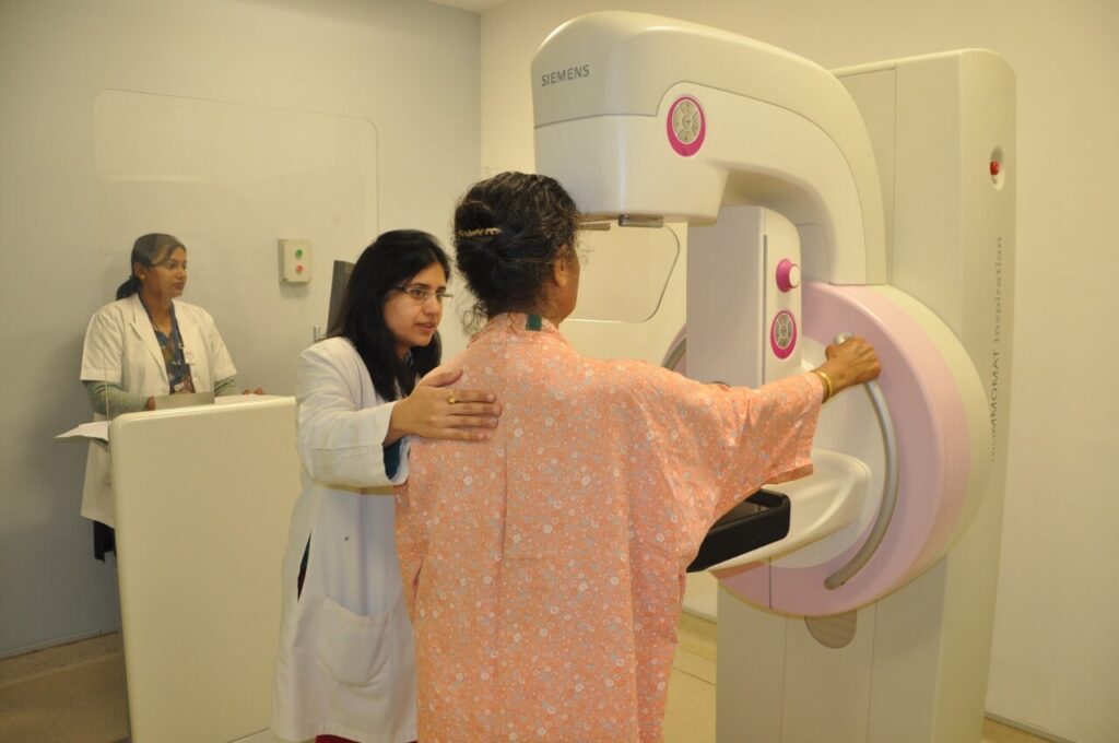
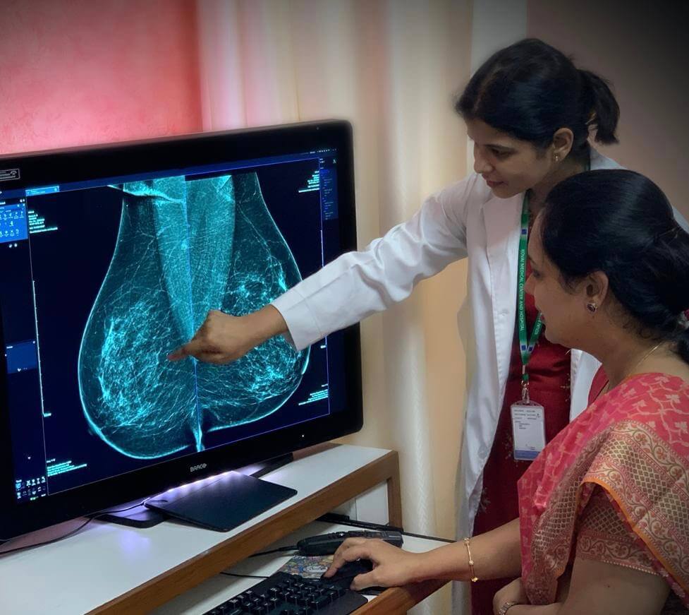
Mammography is the examination of the breast using low-dose x-rays. Two views are taken for each breast. Mammograms may be the conventional film-based type, which utilizes x-rays to produce an image of the breast on film, or digital type that also uses x-rays but captures the image on a computer, where it can be viewed and altered for optimal contrast. While finding no significant difference between the two techniques when applied to the general population and for most women over 50, studies have shown that digital mammograms detected more cancers in women under 50, women with increased breast density, and premenopausal women.

It is advisable to perform mammography soon after your menstrual cycle as this is known to minimize the discomfort that may occur. Application of deodorant/antiperspirant/talcum powder is to be avoided on the day of the study as the particulate matter in these products may alter the findings.


A screening mammogram  (in women without symptoms) is performed to detect early breast cancer when it is completely treatable. It not only help save a life but also save breasts.

In symptomatic women, it is the imaging investigation of choice, especially if no screening mammography has been performed. In young symptomatic females, ultrasound is the first investigation of choice.

What is BI-RADS?

The American College of Radiology (ACR) has established a uniform way for radiologists to describe mammogram findings. The system, called BI-RADS, includes seven standardized categories, or levels. Each BI-RADS category has a follow-up plan associated with it to help radiologists and other physicians appropriately manage a patient’s care.

BI-RADS also includes four categories of breast density that may be reported. The radiologist who reads the mammogram chooses the category that best describes the level of breast density seen on the mammogram film.
​The categories, from the least amount of breast density to the highest, are as follows:

  • The breasts are almost entirely fatty
  • There are scattered areas of dense glandular tissue and fibrous connective tissue (together known as fibroglandular density)
  • The breasts are heterogeneously dense, which means they have more of these areas of fibroglandular density. This may make it hard to see small masses in the breast tissue on a mammogram.
  • The breasts are extremely dense, which makes it hard to see tumors in the breast tissue on a mammogram.

What is the best method of screening for breast cancer?

Regular high-quality screening mammograms and clinical breast exams are the most sensitive ways to screen for breast cancer.

Regular breast self-exam, or BSE—that is, checking one’s own breasts for lumps or other unusual changes—is not specifically recommended for breast cancer screening. In clinical trials, BSE alone was not found to help reduce the number of deaths from breast cancer.

However, many women choose to examine their own breasts. Women who do so should remember that breast changes can occur because of pregnancy, aging, or menopause; during menstrual cycles; or when taking birth control pills or other hormones. It is normal for breasts to feel a little lumpy and uneven. Also, it is common for breasts to be swollen and tender right before or during a menstrual period. Whenever a woman notices any unusual changes in her breasts, she should contact her health care provider.

What is 3-D Mammography (3D Tomography)?

Three dimensional (3-D) mammography, also known as digital breast tomosynthesis, is a type of digital mammography in which low dose x-rays are used to capture images of the breast from different angles and then with the help of computer software, this data is reconstructed into images of slices through the breast thickness. It is similar to how the compute tomography (CT) scanner is used to create images through the body.

It also utilises low dose x-rays , however, till date, its use is recommended only along with the standard 2-D digital mammography, so that the cumulative dose is higher compared to the 2-D mammography done alone but it is still within the accepted limits. Currently, the studies and researches are going on to replace the 2 D mammograms with the synthesized 2D digital mammograms obtained from the tomosynthesis data. This will help in lowering down the radiation dose.