


Dr. Varshaa Hardas
Star Imaging And Research Centre
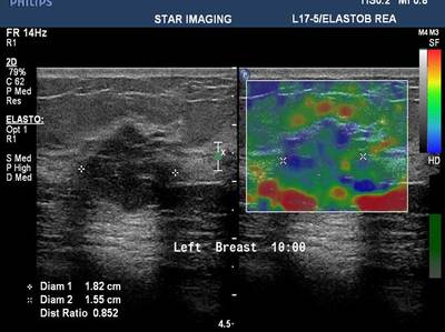
39yr old lady with history of lump in the left breast UIQ, No previous imaging done. No positive family history for CA Breast/Ovary. Pre-Menopausal Status.
Findings
Mammography- Fairly well circumscribed predominantly sold lesion in the left breast UIQ with partially obscured margins. No spiculations. No associated microcalcifications noted. Sonography: Well defined hypoechoiec lesion with smooth margins.
Answer
Low-grade Mucinous Carcinoma ER?PR- +ve HER-2 NEU -VE
Together we are making a difference- and, you can too..
Breast Imaging Society, India was founded by a group of dedicated breast radiologists in 2013 with the idea to improvise and standardize breast imaging in India.