


Dr Jeevanjot Matharoo
Medanta, The Medicity, Gurgaon, Haryana
A 45 year old lady presented with persistent bloody nipple discharge from right breast since 1 month.
Non calcified Ductal Carcinoma In Situ
Discussion
Ductal carcinoma in situ (DCIS) is a noninvasive malignancy and a nonobligate precursor to invasive cancer . Although the detection of DCIS has increased with the advent of widespread mammography screening, it is essential to have more accurate assessment of the extent of DCIS for successful breast conservation surgery. (1) DCIS most commonly appears as microcalcifications at mammography which may be amorphous, coarse, heterogeneous, or fine pleomorphic with a clustered, linear, or segmental distribution. Majority of the time, microcalcifications are the sole finding of DCIS, although focal asymmetry, diffuse increased reticular densities, focal architectural distortion or masses are also observed. (2) Mammographic determination of the extent of DCIS usually depends on the presence of calcifications. Because calcifications are not present in all cases of DCIS, such lesions are mammographically occult, contributing to a mammographic sensitivity of 70–80% . Similarly, because all involved areas may not calcify equally, the extent of disease is often underestimated on mammography. (3) Therefore, it is possible that mammography would fail to depict prognostically relevant DCIS in a large number of women in case of non calcified DCIS. Contrast-enhanced MRI of the breast is complementary to mammography in the detection of DCIS because enhancement may be seen in areas of calcified as well as noncalcified DCIS. Therefore, MRI is helpful in detection of noncalcified disease and more accurate assessment of the disease extent, improving treatment and prognosis. (4) DCIS most commonly manifests as non mass enhancement in segmental or linear/ ductal distribution with internal enhancement patterns like clumped, heterogeneous and clustered ring enhancement. In case with negative mammogram which may harbor DCIS but demonstrated by MRI, it remains important to be aware that high grade DCIS may present as a non‐calcified mammographic abnormality. High‐grade DCIS appears to be more easily detected than low‐grade, suggesting MRI may have a significant benefit in excluding high‐grade disease with a negative mammogram.
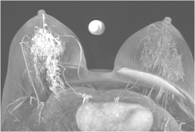
Findings
Bilateral Mammograms revealed increased reticular densities in the upper half of the right breast. A 16 mm well defined nodule seen in the left subareolar region. BIRADS 3/4 Bilateral breast ultrasound revealed extensively dilated ducts showing thickened walls and intra ductal contents corresponding to the increased reticular densities on the mammogram of right breast. Left breast revealed a 17 x 10 mm well defined low echogenic lesion with posterior enhancement in the left subareolar region. BIRADS 3 MRI showed segmental clumped / clustered ring enhancement extending from 10 to 2 O’ clock position in the right breast with extensive ductal branching and intraluminal contents extending till the nipple suspicious for DCIS. Left breast revealed a benign 16 x 14mm well defined nodule in the lower inner quadrant close to the nipple. BI-RADS-4.
References
Heather I. Greenwood, Samantha L. Heller et al.Ductal Carcinoma in Situ of the Breasts: Review of MR Imaging Features. RadioGraphics 2013; 33:1569–1588 2) Takayuki Yamada, Naoko Mori et al.Radiologic-Pathologic Correlation of Ductal Carcinoma in Situ. RadioGraphics 2010; 30:1183–1198 Jennifer H. Menell, Elizabeth A. Morris. Determination of the Presence and Extent of Pure Ductal Carcinoma in Situ by Mammography and Magnetic Resonance Imaging. The Breast Journal 2005, 11 (6): 382–390 Christiane K. Kuhl.Science to Practice: Why Do Purely Intraductal Cancers Enhance on Breast MR Images? Radiology2009: 253(2) :281-283
Together we are making a difference- and, you can too..
Breast Imaging Society, India was founded by a group of dedicated breast radiologists in 2013 with the idea to improvise and standardize breast imaging in India.